Gladstone NOW: The Campaign Join Us on the Journey✕


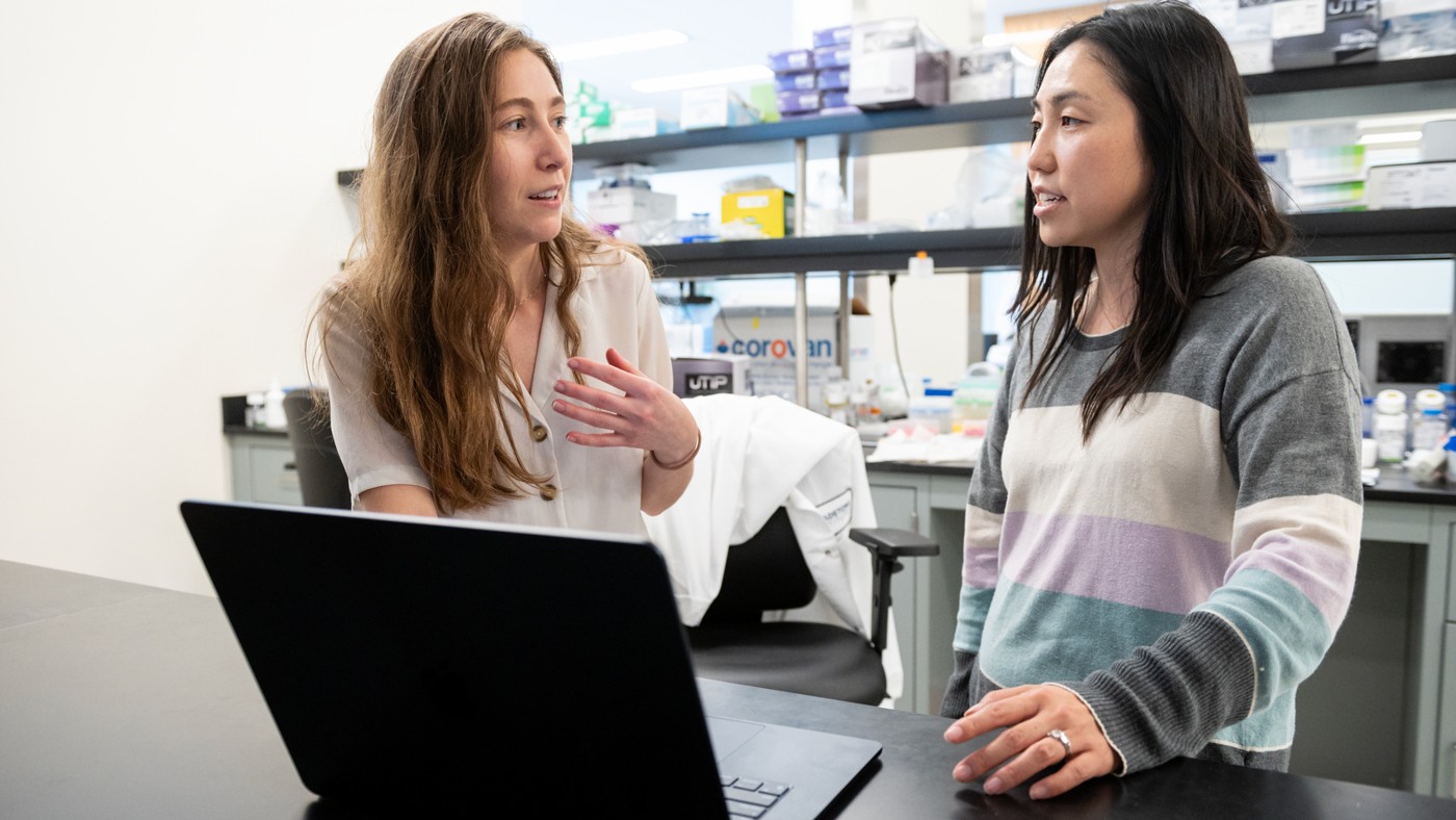
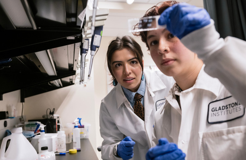
A novel method developed by Gladstone researchers is already answering longstanding questions about basic DNA dynamics—and may one day lead to new ways of treating cancer and other conditions. Seen here are co-authors Nour Abdulhay (left), a former graduate student in the Ramani Lab, and Laura Hsieh (right).
Within each of the trillions of cells in our bodies, more than six feet of DNA is carefully packaged, encoding instructions for the biological processes we need to live. The secret to that precise packaging, which allows so much DNA to compress into the tiny space of a cell’s nucleus, lies in the power of the nucleosome.
Like beads on a string, nucleosomes are tight bundles of DNA that occur along the length of a DNA strand, helping condense genetic information until it’s needed for a specific biological function.
Importantly, nucleosomes restrict access to the instructions encoded by the wound-up DNA, hindering expression of the concealed genes. Regulatory proteins shuttle the nucleosomes back and forth along DNA strands, winding or unwinding different stretches of DNA to expose or conceal them—switching certain functions on or off.
But exactly what drives this intricate process has remained a mystery, with competing models explaining how regulatory proteins rearrange nucleosomes along DNA strands. Until now.
Researchers at Gladstone Institutes have developed a new, high-precision tool that enabled them to identify the correct model. Their findings, published in the journal Nature Structural & Molecular Biology, show that the regulatory proteins arrange nucleosomes so they are spaced out along a DNA strand, instead of cinched closely together.
“This tool resolved an important question about the fundamental dynamics of DNA packaging, with potential relevance for many different human diseases,” says Vijay Ramani, PhD, assistant investigator at Gladstone and senior author of the study. “There are many more mysteries we could solve with the same approach—we see this is a great initial illustration of its power.”
Spaced Out, Not Cinched Up
Our cells contain several different types of proteins that operate in unique ways to rearrange nucleosomes and manage which cellular processes are turned on or off at any moment. For this study, Ramani and his colleagues focused on a specific family of regulatory proteins known as independent switch (ISWI) chromatin remodelers.
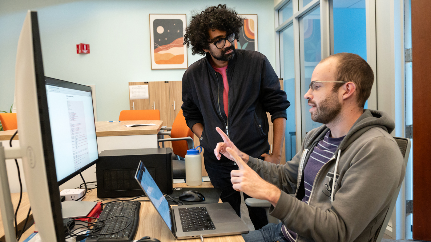
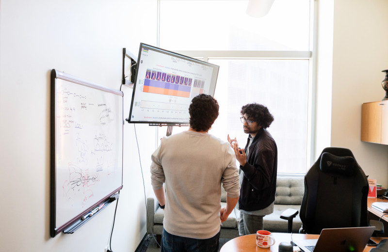
Gladstone Assistant Investigator Vijay Ramani (left) speaks with Colin McNally (right), who helped develop a novel tool called Chromatin Accessibility on Assembled Templates, or ChAAT.
“Mutations in ISWI proteins have been linked in some way to more than one third of all types of cancer, and to multiple neurodevelopmental disorders and other diseases,” says Ramani, who is also an assistant professor of biochemistry and biophysics at UC San Francisco (UCSF). “So, it is really important to understand how they operate.”
Prior research has resulted in two competing models for how ISWI proteins might rearrange nucleosomes. In the “clamping” model, the proteins cinch nucleosomes together, like sliding all the beads on an abacus to one side. In contrast, the “length-sensing” model has nucleosomes positioned like beads spaced out along an abacus, with the distance between them varying depending on their density.
Geeta Narlikar, PhD, professor of biochemistry and biophysics at UCSF and co-author of the new study, developed the length-sensing model several years ago based on biophysical measurements made in her lab. But other labs have reported measurements that support the clamping model.
“The problem is that, to date, the methods available to measure nucleosome positioning require cutting multiple DNA strands into many small fragments, and then averaging your measurements across fragments,” Ramani says. “You can imagine how that would only give a vague idea of how nucleosomes are positioned with respect to each other.”
A Tool That Builds Instead of Destroys
To finally settle the score between the two models, the researchers needed a two-part strategy. First, they had to be able to take precise measurements of nucleosome spacing along a single DNA strand without first destroying it. Ramani’s team, including the study’s lead author Nour Abdulhay, had already developed a protocol to do just that—a tool they call the single-molecule adenine methylated oligonucleosome sequencing assay (SAMOSA).

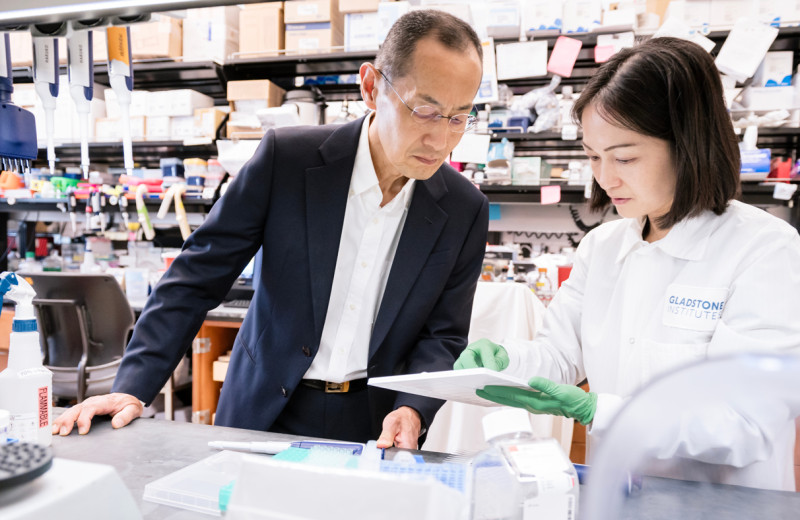
Gladstone Research Associate Megan Ostrowski (left), a member of the research team, speaks with Assistant Investigator Vijay Ramani (right), senior author of the new study.
Second, they needed to be sure they were capturing the specific actions of ISWI proteins, without any other influences.
“The cell is a beautiful machine, but it's an incredibly intricate and complex one,” Ramani says. “If you really want to understand the behavior of a certain group of proteins on a DNA-protein substrate, you need to reconstitute the system, you need to build it up like an engineer would.”
So, Ramani and his team developed a new companion tool for SAMOSA called Chromatin Accessibility on Assembled Templates (ChAAT), which enables them to essentially build a DNA strand with associated nucleosomes from scratch, and then add ISWI proteins to study their effects. (Adding a bit of levity to Ramani’s serious work, he’s named both tools after some his favorite Bombay snacks—an homage to his Indian heritage.)
Using SAMOSA-ChAAT in a series of experiments, the team precisely measured the distances between nucleosomes on DNA strands in the context of ISWI protein activity. They found that distances varied depending on the number of nucleosomes on a given stretch of DNA, rather than always being spaced at a consistent, closely-set distance. This evidence suggests that the length-sensing model, not the clamping model, most accurately captures how ISWI proteins work.
“We went even further by doing computational simulations to be absolutely sure that the analyses we used for the experiments would have detected the clamping model if it really was happening,” Ramani says. “In this way, we integrated alternative modes of thinking about the problem before settling on the length-sensing model as the correct one.”
By confirming the length-sensing model, this work could pave the way to a better understanding of the many diseases in which ISWI protein function is disrupted—perhaps one day leading to better treatments.
The team anticipates that SAMOSA-ChAAT could be applied to deepen understanding of many other processes involving DNA-nucleosome structures, which are collectively known as chromatin.
“There are so many opportunities because all of the essential processes that act on DNA—including replication, transcription, and repair—do so in the context of chromatin dynamics,” Ramani says. “We think that SAMOSA-ChAAT opens up a frontier of discovering totally new insights into these processes, and we’re excited to tackle whatever we can with it.”
For Media
Kelly Quigley
Director, Science Communications and Media Relations
415.734.2690
Email
About the Study
The paper, “Nucleosome density shapes kilobase-scale regulation by a mammalian chromatin remodeler,” was published in the journal Nature Structural & Molecular Biology on September 11, 2023.
Other authors are Colin P. McNally, Megan S. Ostrowski, Camille M. Moore, Arjun S. Nanda, and Ke Wu of Gladstone; Laura J. Hsieh, Un Seng Chio, Ziling Zhou, and Hani Goodarzi of UCSF; Mythili Ketavarapu of UC Santa Barbara; and Sivakanthan Kasinathan of Lucille Packard Children’s Hospital, Stanford University.
This work was supported by the National Institutes of Health (NIH; DP2-HG012442, U01-DK127421, R35-GM127020), the UCSF Program for Breakthrough Biomedical Research, and the UCSF Sandler Fellows program.
About Gladstone Institutes
Gladstone Institutes is an independent, nonprofit life science research organization that uses visionary science and technology to overcome disease. Established in 1979, it is located in the epicenter of biomedical and technological innovation, in the Mission Bay neighborhood of San Francisco. Gladstone has created a research model that disrupts how science is done, funds big ideas, and attracts the brightest minds.
Featured Experts
Support Discovery Science
Your gift to Gladstone will allow our researchers to pursue high-quality science, focus on disease, and train the next generation of scientific thought leaders.
A Gene That Keeps Stem Cells From Losing Their Way
Article
April 30, 2026
A Gene That Keeps Stem Cells From Losing Their Way
Scientists find a gene essential for keeping intestinal stem cells stable, offering insight into how tissues repair themselves.
News Release Research (Publication) Yamanaka Lab Stem Cells/iPSCsGladstone Recognized in Inaugural Cure Innovation Index for Translating Science Into Real-World Health Impact
Article
April 30, 2026
Gladstone Recognized in Inaugural Cure Innovation Index for Translating Science Into Real-World Health Impact
Gladstone ranks 15th among 60 institutes and centers nationally for its ability to turn discoveries into measurable health outcomes.
Institutional News News ReleaseAI Discovery Reveals DNA Isn’t Locked Away in Cells After All
Article
April 29, 2026
AI Discovery Reveals DNA Isn’t Locked Away in Cells After All
A new study upends long-held view about how DNA is stored, explaining how subtle shifts in gene activity contribute to cancer, aging, and other complex diseases.
News Release Research (Publication) Cancer Data Science and Biotechnology Ramani Lab Aging AI