Gladstone NOW: The Campaign Join Us on the Journey✕


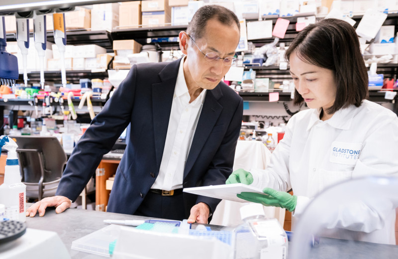
A team led by Nevan Krogan at Gladstone and UCSF has demonstrated that a large-scale and systematic genetic approach can indeed yield reliable and detailed information on the structure of protein complexes. Photo: QBI, UCSF.
One of biologists’ most vexing tasks is figuring out how proteins, the molecules that carry the brunt of a cell’s work, do their job. Each protein has a variety of knobs, folds, and clefts on its surface that dictate what it can do. Scientists can visualize these features fairly easily on individual proteins. But proteins don’t act alone, and scientists also need to know the shape and composition—the structure, as they call it—of the complexes that proteins form when working together.
With precise information about the structure of protein complexes, scientists stand better chances of designing efficient drugs to block or boost the complexes’ activity for therapeutic applications. They can also better anticipate how a mutation might disrupt a complex and lead to disease.
But determining the structure of protein complexes is a painstaking endeavor. Every complex is different, there is no one-size-fits-all way to determine their structure, and few means to speed up the process. Most importantly, the methods that yield the most precise structural information entail taking the complexes out of their natural context—the cell. As a result, scientists peering at a structure are faced with a nagging doubt: Does it really reflect how the complex looks and works when it’s still in the cell?
Ultimately, proteins come from genes, and since genes have proven easier to work with than proteins, some scientists are looking to genes and a fast-growing arsenal of genetic tools to facilitate the determination of protein structures.
Now, a group at Gladstone Institutes and UC San Francisco (UCSF) has demonstrated that a large-scale and systematic genetic approach can indeed yield reliable and detailed information on the structure of protein complexes. Their findings are published in the journal Science.
“Our technique allows us to collect large amounts of structural data from live cells, reflecting how the proteins work in their normal environment rather than in artificial lab conditions,” says Nevan Krogan, PhD, who led the study and is a senior investigator at Gladstone, as well as a professor of cellular and molecular pharmacology and the director of the Quantitative Biosciences Institute (QBI) at UCSF. “This has not been possible on such a scale before, and it should greatly speed up the process of determining the structure of protein complexes, including those that are difficult to tackle with traditional methods.”
The approach builds on a technology that Krogan pioneered called genetic interaction mapping. It screens through thousands of combinations of gene mutations, in live cells and in relatively little time, and can reveal genes whose protein products work in common cellular processes. Krogan and his team cranked up the resolution power of these screens and successfully modeled two protein complexes in yeast cells, and one in bacterial cells.
Krogan sees this advance not as a replacement for other ways of determining the structure of proteins, but as a crucial complement, part of a strategy called “integrative modeling” pioneered by Andrej Sali, a professor at UCSF and a collaborator on this project.
“Combining the genetic data from our screens with other structural information improved the accuracy of our models,” Krogan says. “Our study highlights the power of integrative modeling and the value of combining several data sets gathered in completely different ways.”
From Yeast to Human Cells and Diseases
Proteins are chains of building blocks called amino acids. Deciphering the structure of a protein complex consists mainly in figuring out which stretches of amino acids end up close to one another when the complex is assembled. Most of the time, this is achieved through biochemistry.
Instead, Krogan and his team relied on genetics, and looked at how the amino acids of a complex behaved in their large-scale screens. The idea is that if two amino acids are close to each other—say, within the same knob or cleft at the complex’s surface—they are likely to perform similar functions for the complex. Therefore, in a genetic screen, the two amino acids are expected to interact with the same genes. But protein complexes are indeed complex, with different regions potentially influencing each other or performing similar functions.
“So, if two amino acids in a complex interact with the same gene, they may or may not be close to each other,” says Hannes Braberg, PhD, co-first author of the study and a scientist at QBI, an organized research unit under the School of Pharmacy at UCSF. “But if they interact with the same 50 genes out of 1,000 possibilities, then the chances are much greater that they are indeed close to each other in the complex.”
The scientists decided to explore whether this reasoning could be used to determine the structure of protein complexes in their native environment—live, growing cells.
They started with two proteins called Histone H3 and Histone H4, which form a well-understood protein complex. They performed their screen in yeast cells and used the resulting information to model the structure of the histone H3-H4 complex.
“The structure we obtained was consistent with existing data about the protein complex,” says Braberg. “And the performance of our method is comparable to that of a commonly used biochemistry approach, which is remarkable, given that the genetic interaction data is purely based on looking at how well cells grow!”
The success of their approach was not limited to the H3-H4 complex, as the researchers obtained similar results with two other protein complexes, one in yeast and one in bacterial cells. This bodes well for the widespread application of the approach to many more complexes, particularly complexes that do not yield easily to traditional techniques because, for instance, they are embedded in other cellular components, or are too large or too short-lived.
“Recent advances in CRISPR-Cas9 genome editing should also enable us to extend our approach to human cells,” says Krogan. “This possibility opens exciting perspectives to investigate diseases caused by gene mutations or pathogens.”
Combining CRISPR with genetic interaction screens, Krogan and his team will be able to precisely describe the impact of disease mutations on the structure of protein complexes, and identify the changes that are relevant to disease. His team recently used genetic interaction screens to study the interface between viruses and human cells. Building on this work, they can now introduce specific mutations into the genome of a pathogen, and use genetic interaction profiles of the human host proteins to understand the consequences on infection in live cells.
“This project, using genetic interaction screens to inform the structural understanding of protein complexes, started 15 years ago,” Krogan says. “We have continued refining the approach and increasing its power over the years, and it’s really gratifying to see the unprecedented resolution with which it can now inform us about biological phenomena as they take place inside live cells.”
For Media
Julie Langelier
Associate Director, Communications
415.734.5000
Email
About the Study
The paper, “Genetic interaction mapping informs integrative structure determination of protein complexes,” was published online by Science on December 10, 2020.
Other co-first authors are Stefan Bohn from Gladstone Institutes, and Ignacia Echeverria and Peter Cimermancic from UCSF. Other authors include Jiewei Xu, Michael Shales, Ruth Hüttenhain, Shuyi Wang, David Mavor, Riccardo Pellarin, Dina Schneidman, James S. Fraser, Derek Bogdanoff, Kaitlin K. Chaung, John Morris, Anthony Shiver, Richard Alexander, Carol Gross and Andrej Sali from UCSF; Shuangying Jiang and Junbiao Dai from Shenzhen Institutes of Advanced Technology, Chinese Academy of Sciences, China; Gajendradhar Dwivedi and James E. Haber from Brandeis University; Joel S. Bader and Jef D. Boeke from Johns Hopkins University School of Medicine; and Raghuvar Dronamraju and Brian D. Strahl from University of North Carolina School of Medicine.
The work was funded by the National Institutes of Health, the National Key Research and Development Program of China, and the National Natural Science Foundation of China.
About Gladstone Institutes
Gladstone Institutes is an independent, nonprofit life science research organization that uses visionary science and technology to overcome disease. Established in 1979, it is located in the epicenter of biomedical and technological innovation, in the Mission Bay neighborhood of San Francisco. Gladstone has created a research model that disrupts how science is done, funds big ideas, and attracts the brightest minds.
Featured Experts

Support Discovery Science
Your gift to Gladstone will allow our researchers to pursue high-quality science, focus on disease, and train the next generation of scientific thought leaders.
A Gene That Keeps Stem Cells From Losing Their Way
Article
April 30, 2026
A Gene That Keeps Stem Cells From Losing Their Way
Scientists find a gene essential for keeping intestinal stem cells stable, offering insight into how tissues repair themselves.
News Release Research (Publication) Yamanaka Lab Stem Cells/iPSCsGladstone Recognized in Inaugural Cure Innovation Index for Translating Science Into Real-World Health Impact
Article
April 30, 2026
Gladstone Recognized in Inaugural Cure Innovation Index for Translating Science Into Real-World Health Impact
Gladstone ranks 15th among 60 institutes and centers nationally for its ability to turn discoveries into measurable health outcomes.
Institutional News News ReleaseAI Discovery Reveals DNA Isn’t Locked Away in Cells After All
Article
April 29, 2026
AI Discovery Reveals DNA Isn’t Locked Away in Cells After All
A new study upends long-held view about how DNA is stored, explaining how subtle shifts in gene activity contribute to cancer, aging, and other complex diseases.
News Release Research (Publication) Cancer Data Science and Biotechnology Ramani Lab Aging AI