Disrupted Boundary Between Cell Types Linked to Common Heart Defects
Listen:
Every year, nearly 40,000 U.S. babies—or about one in 100—are born with a hole in the wall between their heart’s chambers. For many of them, it can mean a lifetime of surgeries, medications, and careful monitoring.
These congenital heart defects are the most common type of birth defect, yet scientists have struggled to understand exactly how this critical wall of muscle, called the interventricular septum, forms during development and why it so often goes wrong.
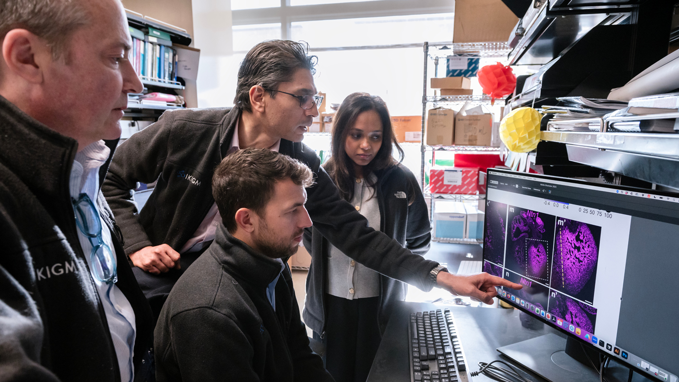
Now, researchers at Gladstone Institutes and UC San Francisco (UCSF) have discovered that a boundary between two populations of heart cells, established very early in development, acts as a crucial guide for proper heart formation. When this boundary is disrupted, cells mix inappropriately, leading to holes in the interventricular septum and other defects, according to a new study featured on the cover of the January issue of Nature Cardiovascular Research.
“We’ve uncovered a completely new mechanism for how the heart patterns itself during development,” says Benoit Bruneau, PhD, director of the Gladstone Institute of Cardiovascular Disease and co-senior author of the new study. “Our study offers brand-new insight into the incredible degree of finesse required to assemble an organ like the heart.”
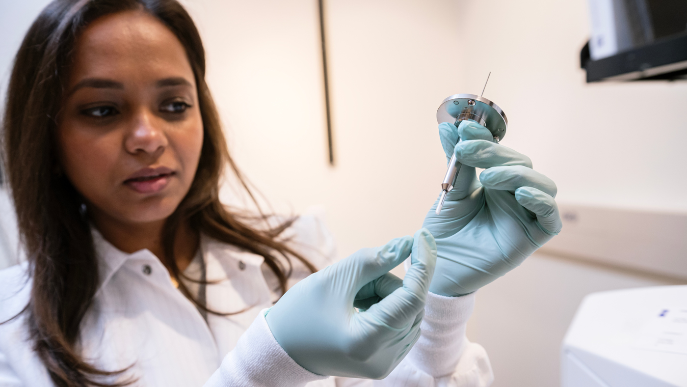
To date, scientists have identified hundreds of genes involved in heart development, but understanding exactly how each one contributes is challenging. But nearly half of all heart defects still have no known genetic cause.
“By further studying the pathways that we’ve identified here, we hope to eventually pinpoint some of those causes and identify new potential therapies,” says Irfan Kathiriya, MD, PhD, professor in the Department of Anesthesia and Perioperative Care at UCSF and co-senior author of the new study.
A Gene That Causes Heart Defects
One gene in particular has puzzled researchers for decades: TBX5. Mutations in this gene cause Holt-Oram Syndrome, a condition that includes holes in the interventricular septum—but no one understood why or how.
“I’ve been studying this gene for over 25 years, trying to understand how it causes heart defects,” says Bruneau, who is also William H. Younger Chair in Cardiovascular Research at Gladstone and a professor of pediatrics at UCSF. “My own daughter was born with this type of defect, so understanding these mechanisms is deeply personal to me.”

To solve this mystery, Bruneau teamed up with Kathiriya. They tracked cells in which the gene TBX5 was active during heart development in mice.
They expected cells with TBX5 turned on—usually found on the heart’s left side—to be separate from cells expressing another gene, MEF2C, found on the right side. Instead, they found a thin stripe of cells with both genes turned on, located at the future site of the interventricular septum, between the heart’s left and right ventricles.
“We were really surprised to see this and it made us think of a concept in developmental biology called a compartment boundary,” says Bruneau.
Maintaining Boundaries
A compartment boundary, he explains, isn’t a physical wall, but rather a population of cells with special properties that actively prevent neighboring cell populations from mixing. Few such boundaries have been discovered in mammals.
The researchers watched the doubly-marked cells in more detail over the course of development. They found that the stripe of cells appears before the heart even forms and that it maintains its shape and position as the organ develops. When they destroyed the boundary cells early in development, something striking happened.
“We saw an incredibly disorganized septum, and cells that are normally kept separate were mixing together,” says Kathiriya, who is also a visiting scientist at Gladstone. “It confirmed that these cells aren’t just sitting there; they’re actively maintaining the separation between the left and right sides of the heart.”
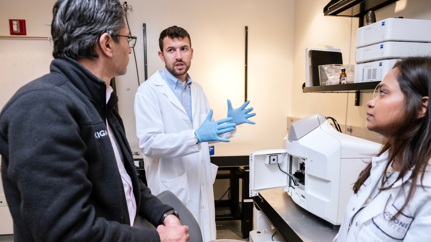
Next, the team reduced levels of the TBX5 gene in developing mouse hearts, mimicking what happens in Holt-Oram Syndrome. The compartment boundary was disrupted—cells were in the wrong position, lost their organized structure, and the cells usually found on the left and right sides of the heart were mixed.
“Instead of being arranged like a Roman phalanx with shields all aligned, now the soldiers were disoriented, facing the wrong way,” Bruneau explains. “They’d lost their commander.”
The experiments confirmed that TBX5 is responsible for maintaining the boundary compartment separating the two sides of the heart.
Guidance Molecules Point the Way
TBX5 itself is a transcription factor—a type of protein that controls hundreds of other genes—making it difficult to target with drugs. So, the scientists analyzed which other genes were turned on and off in the newly identified boundary compartment cells.
They discovered that TBX5 controls the levels of two other molecules, SLIT2 and Netrin-1, that were already well known for their roles in brain development, where they help neurons find their correct positions. But neither protein had been implicated in the heart before.
“In the brain, these molecules guide cells to the right place,” Kathiriya says. “We found they’re doing something remarkably similar in the heart.”
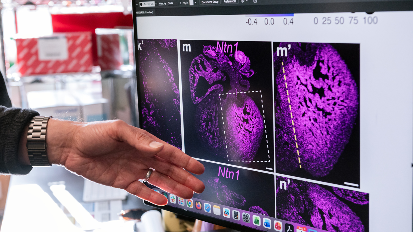
Mice lacking either of these molecules developed disrupted compartment boundaries and holes in their interventricular septum. Moreover, when the researchers adjusted levels of Netrin-1 in mice with TBX5 mutations, they partially prevented the heart defects. The finding could point toward new drugs for preventing or treating Holt-Oram Syndrome or other heart defects caused by a malformed boundary compartment.
“Unlike transcription factors like TBX5, signaling molecules like SLIT2 and Netrin-1 are targets we can potentially manipulate with drugs,” Kathiriya says. “These pathways are already being targeted in cancer and other diseases.”
The researchers plan to continue studying how these molecules work in the developing heart, hoping to identify additional genetic causes of birth defects and therapeutic targets.
Up Next


American Heart Association Honors Benoit Bruneau with 2026 Distinguished Scientist Award
Bruneau, director of the Gladstone Institute of Cardiovascular Disease, will receive the association’s highest scientific honor in November.
Misfolded DNA Blueprint: A New Origin for Genetic Disease
Gladstone researchers found losing just one copy a particular gene can disrupt heart development and lead to congenital heart disease.
Scientists Pinpoint a Key Gene Behind Heart Defects in Down Syndrome
After decades of mystery, Gladstone researchers identify a gene that can derail heart formation—and show that fixing it prevents the problem in mice.
CIRM Awards $7.5 Million in Discovery Grants to Gladstone Investigators
Two ambitious research projects led by Gladstone investigators are boosted by funds from the California Institute for Regenerative Medicine.

Get Involved
Join the Team
Our people are our most important asset. We offer a wide array of career opportunities both in our administrative offices and in our labs.
Open PositionsPartner With Us
Discoveries don’t stay in our labs. Gladstone facilitates spinout companies to advance promising therapies, shortening the path between scientific discovery and helping real patients.
Learn MoreGive
Your gift to Gladstone will allow our researchers to pursue high-quality science, focus on disease, and train the next generation of scientific thought leaders.
Donate Now