1. SCIENCE
  2. CORE FACILITIES
  3. Histology and Light Microscopy

Histology and Light Microscopy

The Histology and Light Microscopy Core provides technical assistance, training, consultation, and collaboration for all aspects of experimental design, sample preparation, image processing, and data analysis.

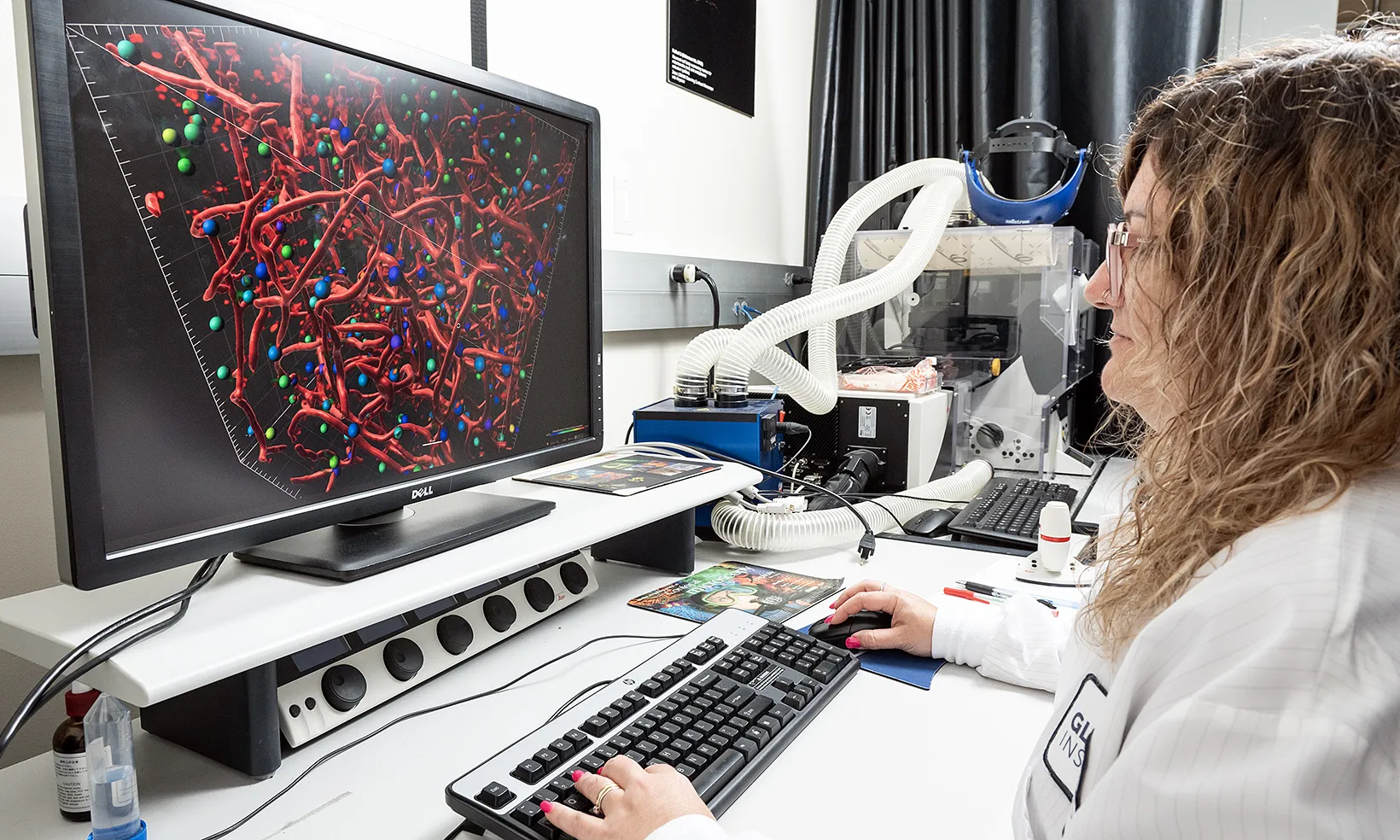
Our core is equipped with state-of-the-art technologies and expertise in histology, high-resolution histological imaging, confocal microscopy, light sheet microscopy.

We strive to provide you with the knowledge and equipment you need to successfully perform your experiment. Our core also offers self service access for trained users.

Services

In addition to technical assistance and consultation on our services, we also provide training so that you can utilize the equipment on your own time.

  • Tissue processing (paraffin/FFPE and frozen/cryo)
  • Tissue embedding
  • Tissue sectioning
  • Tissue staining and whole slide scanning
    • H&E
    • IHC, IFA (Coming Soon)
    • ISH, TUNEL
    • Special staining (PAS, ORO, Congo Red, Masson’s Trichrome)
    • Spatial Biology: 10x Visium

On live and fixed samples

  • Confocal/super-resolution
  • Light-sheet, tissue clearing, volumetric imaging
  • Automated whole slide scanning
  • Widefield microscopy
  • Mammoth: Imaris Workstation (Room 327)
    • Imaris Software
    • Imagescope, ImageJ/FIJI, OlyVia
  • Gladstone Institute of Cardiovascular Disease Workstation

Fees and Scheduling

The Histology and Light Microscopy Core recharges researchers at an hourly rate for training and support with microscopes and sample preparation. Consult with our staff before preparing samples to ensure your reagents match the available equipment.

You are required to complete an initial training session before using any equipment in the core.

If you are part of UC San Francisco:

If you are part of a nonprofit institution or a private company:

Microscope Reservation

Equipment

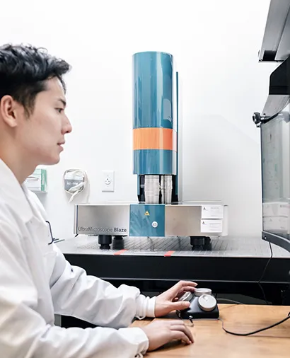
Miltenyi Blaze Ultra-Imaging 3D Light Sheet Microscope

Miltenyi Blaze Ultra-Imaging 3D Light Sheet Microscope

Light Sheet Microscope:

  • Immersion objectives: 1.1X and 4X (0.6X – 10X total range) Camera: 4.2 Mpx sCMOS (2048 x 2048 pixels)
  • 5 lasers: 405, 488, 561, 639, 785nm

Sample types:

  • Pre-cleared sample (aqueous buffers and organic solvents with RI 1.33 – 1.57)
  • Organoids to adult mouse

Application:

  • Imaging (multiple) small to large samples in 3D with low photobleaching and high relative speed

This machine is not optimized for live imaging or structures that need to be resolved with greater than 10X magnification.

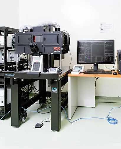
Snow Leopard (Olympus Fluoview FV3000)

Snow Leopard (Olympus Fluoview FV3000)

Specifications:

  • Point-scanning confocal microscope
  • Both Galvo & Resonance scanning for high speed imaging
  • Multiple laser lines (405nm, 445nm, 488nm, 514nm, 594nm, 561nm, 638nm)
  • >Incubation chamber (CO2, Temperature & Humidity) for both short & long live cell/tissue imaging

Sample types

  • Chamber slides, 35mm dishes or optical-quality well plates
  • Both live & fixed tissues/cells

Applications:

  • Scanning confocal imaging for live and fixed -cell, organoid and tissue imaging

This machine is not optimal for very thick samples (>100µm) or very high-speed imaging (>10fps).

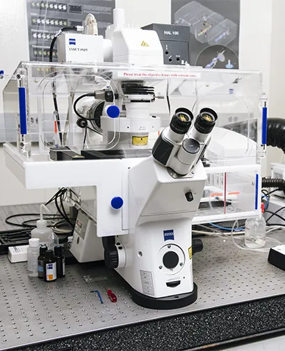
Tarsier (Zeiss LSM880 Super-Resolution AiryScan Confocal Microscope)

Tarsier (Zeiss LSM880 Super-Resolution AiryScan Confocal Microscope)

Specifications:

  • Point-scanning confocal microscope
  • Airyscan detector for increased resolution (>120nm lateral), sensitivity
  • Multiple laser lines (405nm, 445nm, 488nm, 514nm, 561nm, 638nm)

Sample types:

  • Chamber slides
  • 35mm dishes
  • Optical-quality well plates

Applications:

  • Scanning confocal imaging for mid-speed (13 fps) live-cell imaging

This machine is not optimal for very thick samples (>100µm) or very high-speed imaging (>10fps).

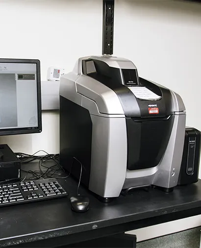
Coyote 1 (Keyence epifluorescence BZX810 )

Coyote 1 (Keyence epifluorescence BZX810 )

Specifications:

  • Automated inverted wide-field epifluorescence and brightfield imaging system
  • Metal halide illumination; filters for DAPI, GFP, RFP, TRITC, and Cy5
  • High sensitivity RGB and monochrome camera

Sample types:

  • Slides or 35mm dishes
  • Culture plates
  • Petri dishes

Applications:

  • Epifluorescence imaging for fixed and live-cell samples

This machine is not recommended for long term live-cell imaging or thick samples (>50um).

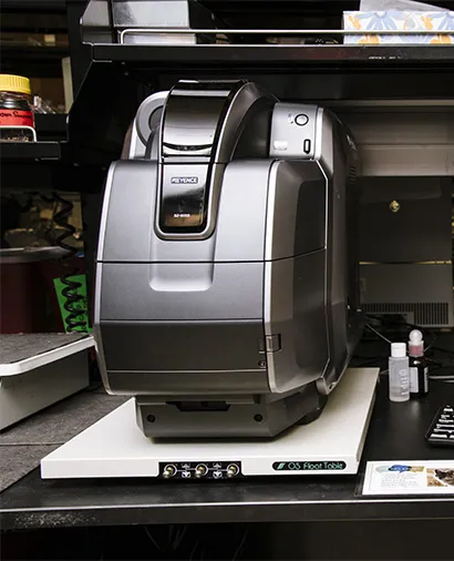
Coyote 2 & 3 (Keyence epifluorescence BZ9000)

Coyote 2 & 3 (Keyence epifluorescence BZ9000)

Specifications:

  • Automated wide-field epifluorescence system
  • Metal halide illumination; filters for DAPI, GFP, RFP, TRITC, and Cy5

Sample types:

  • Slides
  • 35mm dishes
  • Culture plates

Applications:

  • Epifluorescence imaging for fixed and live-cell samples
  • Tiling multiple images for large sample areas

This machine is not recommended for long term live-cell imaging, thick samples (>50um), or plastic tissue culture plates.

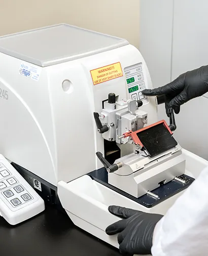
Leica RM2245 Automatic Microtome

Leica RM2245 Automatic Microtome

Specifications:

  • The RM2245 is a semi-motorized rotary microtome, designed for routine in histopathology
  • Manual sectioning is enhanced by a high-precision motorized specimen feed, which results in efficient operation with maximum section quality and reproducibility

Application:

  • Choose between conventional, full-hand-wheel rotation, manual sectioning or “rocking mode”.
  • Motorized sectioning and meets the many requirements of modern laboratories

The core does provide training on this device. 

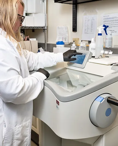
CryoStar NX50 Cryostat Instrument (Epredia)

CryoStar NX50 Cryostat Instrument (Epredia)

Specifications:

  • A form-fitting design, integrated height control
  • Light touch manual handwheel
  • Section thickness from 0.5 µm to 500 µm
  • Vertical stroke length of 64 mm
  • 48 mm horizontal feed range
  • 20 µm specimen retraction on return stroke, with optical indication

Applications:

  • Mounting tissue to ready-to-cut with quick freeze and setup features
  • Sectioning frozen tissue samples

The core provides training on this device. 

FAQs

If you’re a UC San Francisco user, learn how to create an account with iLab.

If you’re part of a nonprofit organization or a for-profit company, you’ll need to sign a legal service agreement (LSA). Once the LSA is signed by both organizations, you’ll receive instructions on how to set up an iLab account to request services. Contact the core to start the LSA process.

  1. Login into your iLab account
  2. Initiate a service request
  3. Use the temporary PO number 11111 to finalize and submit your service request
  4. Email the core after your service request is submitted with the temporary PO to receive a formal quote
  5. Use the quote to generate a formal PO by your lab or organization’s financial department
  6. Use the valid PO to update your iLab account profile and billing or financial information
  7. Email the core a PDF copy of your formal PO

If you need help generating or submitting your PO, reach out to your lab’s grant or financial manager.

When you create an iLab account, you will provide a valid PO. We do not accept credit cards or cash.

The core’s services are available to both Gladstone and outside researchers.

Users can use some of the core equipment once they are trained and granted access by the core’s staff. Only formally trained users are allowed to use our core’s equipment.

To request training, submit an iLab request by clicking on the “request services” tab.

All samples must be dropped off in person. To drop off your samples, stop by the core located in room 367 at Gladstone Institutes, between 10am–3pm.

  • FFPE samples:
    • You should fix your samples (in 4 percent PFA or 10 percent formalin), rinse (3x) in 1x PBS, and store in 70 percent EtOH, before bringing to the core.
    • Gradually increase EtOH ethanol concentration: 25 percent, 50 percent, and finally 70 percent EtOH approach is preferable or recommended.
  • Frozen tissues: Samples can be brought to the core in one of the following conditions:
    • In 30% sucrose (transported on ice)
    • In OCT blocks or tissues slides (transported on dried ice)

Contact the core for the 10x Genomics Visium sample preparation

Sample pick-up
The core will let you know when your samples are ready. When they are ready for pick-up, stop by room 367 at Gladstone Institutes, between 10am–3pm.
Standard turn-around time is typically 15–20 business days from the date the samples are dropped off and required request steps have been completed, not the project submission date via iLab.

Rush order turn-around is 4 business days

Note that these are estimates and turn-around times can be longer due to the complexity and size of the project and our current workload.

Yes, all tissue should be fixed prior to submission. Most tissues do well with fresh perfusion followed by 4 percent PFA or 10 percent formalin fixation overnight in the fridge. Common exceptions are brain and bone samples, which require extended fixation.

We support the Association of Biomolecular Resource Facilities’ (ABRF) Authorship Guidelines.

The existence of core facilities and their ability to obtain financial support depends in part on proper acknowledgment in publications. This is an important metric of the value of most core facilities.

Examples of when you should cite a core could include acknowledgement of:

  • Space
  • Equipment
  • Purchased core reagents/media
  • Services
  • If a core staff member assists you in the design or execution of your experiments

Publications

Members

Rebecca Blandino, PhD
Assistant Core Director

Rose Guerrero
Research Scientist

Alex Liu
Research Technologist II

Jane Srivastava
Scientific Resource Director

Contact

Information relating to POs, quotes, and administrative inquiries.
Email

Technical questions regarding projects.
Email