Gladstone NOW: The Campaign Join Us on the Journey✕

Katie Pollard, PhD, led a study in Nature Methods comparing 25 different maps scientists commonly use to evaluate three-dimensional genomic data. The resulting guide helps researchers choose the right map for their investigations into human development, evolution, and disease.
Think of DNA as a long string, crumpled up within the tiny space of a cell’s nucleus. It might look disordered and bunched, but the way it folds matters. Which sections of DNA strands are buried deep within a clump, which are freely accessible, and which are close to each other is intrinsically tied to the rest of the cell’s function and behavior.
Comparing tightly packed DNA structures called chromatin—examining how they change over time, differ between health and disease, or vary across cell types—also sheds light on fundamental questions in biology, development, and medicine.
In recent years, a few methods have emerged as the ideal ways to map DNA organization; each visualizes data in the form of what’s called a “chromatin contact map.” Yet, it’s been tricky for scientists to compare hundreds or thousands of these maps at once because it involves grappling with difficult statistical problems.
Time and time again, individual research teams come up with their own comparison methods, leading to dozens of different approaches that often produce conflicting results—leaving scientists uncertain about which is best for their specific biological questions. Some methods are better at identifying tiny, subtle changes to DNA structures, while others point toward larger shifts.
Now, a team led by Katie Pollard, PhD, director of the Gladstone Institute of Data Science and Biotechnology, and graduate student Ketrin Gjoni analyzed 25 different methods of comparing chromatin contact maps, with the ultimate goal of giving biologists and data analysts guidance on which to use when. Their results were published in the journal Nature Methods.
We sat down with Pollard and Gjoni to learn about the biggest takeaways from their study.
First, what is a chromatin contact map?
Gjoni: A chromatin contact map is a heat map that shows, for any given genome, which regions of DNA are physically interacting with each other or are close to one another.
These maps let us figure out how DNA is spatially organized within the nucleus of a cell. That organization can then tell us a lot about which genes are being turned on when, or how one small DNA change might be altering the whole architecture of a genome to affect many genes at once.
Why are researchers interested in DNA structure in the first place?
Pollard: Many scientists are becoming more familiar with programs like AlphaFold that predict how proteins fold. They understand that the way proteins fold up into a three-dimensional structure is key to their function, and that a mutation might change a protein’s function by first changing its structure.
It’s the same for DNA—structure matters a lot. Researchers use chromatin contact maps to investigate whether the genome folds differently in various biological contexts, like during development, in disease states, or between species. They want to determine whether these folding changes influence gene expression, replication, or other cellular functions.
We frequently use chromatin contact maps in my lab. For example, we’ve studied how chromatin organization changes during heart development by examining the transition from stem cells to beating heart cells. We’ve also compared how DNA folds differently between humans and chimpanzees, and how this structure changes in diseases such as autism, cancer, and heart defects.

Ketrin Gjioni, a graduate student in the Pollard Lab at Gladstone Institutes, is first author of the new study in Nature Methods.
Researchers use many different methods for comparing chromatic contact maps. Why was it important to study all of these methods head-to-head?
Pollard: We were launching a new project—using machine learning to predict how DNA folds—and needed to figure out the best comparison method to use. We initially planned to evaluate just a few methods to make an informed decision for our own work. But when we saw how much the methods differed, and how they could really lead to different results from each other, it became clear that a comprehensive benchmark was needed. Not just for us, but for the scientific community overall.
What stood out to you in your comparison of these methods?
Gjoni: We evaluated more than 22,000 pairs of chromatin contact maps to gauge how different methods respond to various types of structural changes. It was striking how two methods designed to answer the same general question (such as: “Are these maps different?”) could produce completely different results. We were also surprised that lessons learned in other fields did not translate perfectly to chromatin biology. For instance, the structural similarity index, or SSIM, commonly used in image analysis, performed especially poorly in our benchmark.
Did one method win out over the others?
Gjoni: No, not at all. It really depends on the biological question. The methods are so different, so it’s important to use multiple approaches and compare results, especially when trying to answer biological questions that are complex or nuanced.
For large datasets, we recommend starting with global methods that quickly highlight the most disrupted maps. Then, more specialized tools can be used to examine those maps in greater detail.
We created a summary table in our paper that guides researchers toward what they should consider, as well as an open-source code repository and framework that other scientists can use if they want to run their own analyses. We mostly want researchers to be more informed about the biases and implications of the methods they use.
What’s next for your lab?
Pollard: Now that we have a clearer idea of how well different methods work, we can apply them to interesting questions about disease, evolution, and development. What I’m especially excited about is using our machine learning program to predict how small genetic changes—such as disease-linked mutations—might affect chromatin structure. Once we have these predictions, we can go into cells, make those genetic changes, and test the genome folding to see if our predictions were right.
Ultimately, we hope that by understanding how genome folding changes, we can uncover new insights into genetic diseases, tumor formation, and developmental disorders—and potentially even guide new treatments. Being able to more effectively compare chromatin contact maps is a step toward that insight.
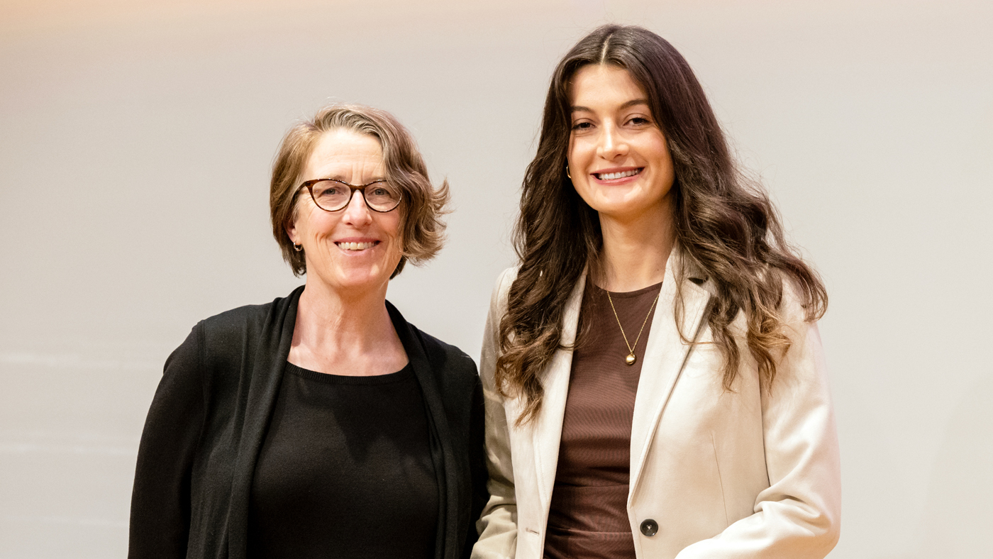
Katie Pollard (left) and PhD candidate Ketjin Gjoni (right) developed practical and useful guidelines that will enable further discovery of the mechanisms of the 3D genome.
For Media
Julie Langelier
Associate Director, Communications
415.734.5000
Email
About the Study
The paper, “Comparing chromatin contact maps at scale: methods and insights,” was published in the journal Nature Methods on March 19, 2025. In addition to Pollard and Gjoni, other authors are Laura Gunsalus, Shuzhen Kuang, and Maureen Pittman of Gladstone; and Evonne McArthur and John A. Capra of UC San Francisco.
The work was supported by the NIH 4D Nucleome Project (U01HL157989), the NIH Office of the Director R03OD034499), the National Institute of General Medical Sciences (R35GM127087, T32GM007347), the National Human Genome Research Institute (F30HG011200), and Additional Ventures.
About Gladstone Institutes
Gladstone Institutes is an independent, nonprofit life science research organization that uses visionary science and technology to overcome disease. Established in 1979, it is located in the epicenter of biomedical and technological innovation, in the Mission Bay neighborhood of San Francisco. Gladstone has created a research model that disrupts how science is done, funds big ideas, and attracts the brightest minds.
Featured Experts
Gladstone NOW: The Campaign
Join Us On The Journey
Disrupted Boundary Between Cell Types Linked to Common Heart Defects
Disrupted Boundary Between Cell Types Linked to Common Heart Defects
Gladstone scientists identified a cellular boundary that guides heart development and revealed how disrupting it can lead to holes in the heart’s wall.
News Release Research (Publication) Congenital Heart Disease Cardiovascular Disease Bruneau LabGene Editing Strategy Could Treat Hundreds of Inherited Diseases More Effectively
Gene Editing Strategy Could Treat Hundreds of Inherited Diseases More Effectively
Scientists at Gladstone show the new method could treat the majority of patients with Charcot-Marie-Tooth disease.
News Release Research (Publication) Neurological Disease Conklin Lab CRISPR/Gene EditingGenomic Maps Untangle the Complex Roots of Disease
Genomic Maps Untangle the Complex Roots of Disease
Findings of the new study in Nature could streamline scientific discovery and accelerate drug development.
News Release Research (Publication) Marson Lab Genomics Genomic Immunology

