Gladstone NOW: The Campaign Join Us on the Journey✕


Li Gan and her team investigated how microglial cells maintain their population.
Microglia, the brain’s immune cells, protect brain cells from infections. They also nurture neurons over their lifespan and prune away excessive connections between neurons. The number of microglia and their distribution in the brain are normally stable—a phenomenon known as homeostasis. However, an excess of activated microglia, called microgliosis, is observed in most neurodegenerative diseases.
Scientists have known for some time that microglia have the remarkable ability to regenerate and restore their population in mouse brains after having been nearly completely wiped out. But the origin of the microglia that repopulate the brain has been a controversial subject. Yet, understanding how microglia normally regenerate could eventually yield new strategies for tackling disease.
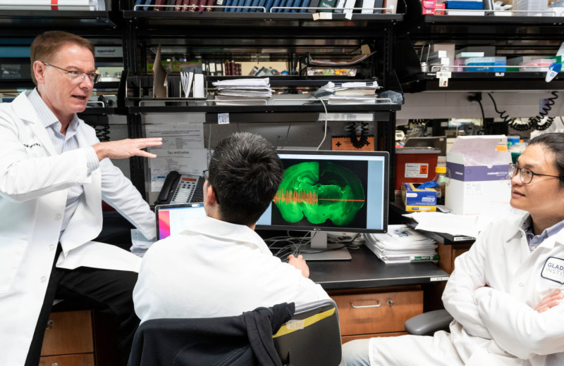
In a recent study published in PLOS Biology, a team of researchers led by Li Gan, PhD, senior investigator at Gladstone Institutes, unlock the mystery of how microglial cells repopulate the brain.
“Our work provides clues to the stability and longevity of microglia,” says Gan. “We hope our findings can open new inroads into the development of therapies for neurodegenerative diseases linked to an excess of microglial cells.”
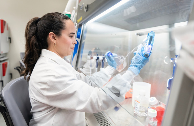
For their study, Gan and her colleagues depleted mouse brains of up to 90 percent of their microglial cells. This allowed them to observe how microglial cells repopulate the brain and eventually restore the network of brain microglia to its original density.
Based on previously published observations, three possible sources for the repopulating microglia have been proposed. They could come from a special pool of brain cells expressing a protein called Nestin1, from a pool of immune cell precursors known as myeloid cells that originate in the bone marrow and circulate in the blood, or from the few microglial cells that remain after depletion.
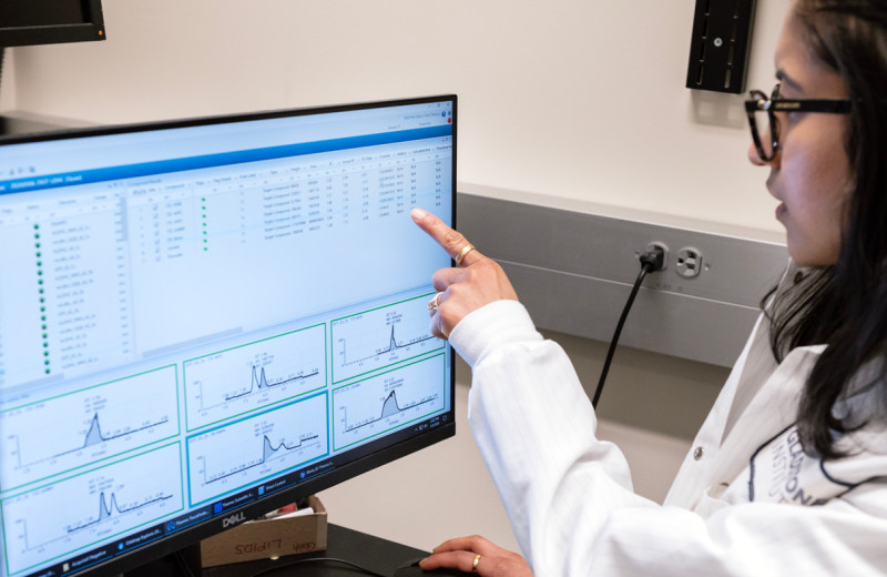
To distinguish among these possibilities, the study’s first author, Lihong Zhan, PhD, used a technique called lineage tracing. With this method, he added a different fluorescent marker to each pool of cells identified as potential sources for regenerating microglia. He then tracked each cell population to identify the one that actually created the new microglial cells.
“We determined, unambiguously, that the vast majority of newborn microglia are generated through self-renewal,” says Zhan, a postdoctoral scholar at Gladstone. “This means the microglia that remained in the mouse brain were responsible for generating the new cells. Our data shows that Nestin1-positive cells and myeloid cells in the periphery contributed very little to the regeneration of microglia.”
Upon initial observation, the regeneration of microglia seemed to resemble disease-associated microgliosis, but eventually resolved to homeostatic level, even after repeated depletion and regeneration.
“We need to continue to investigate how microglial cells know to stop growing and remain within set boundaries in this setting,” says Zhan.
Gan and her team made other interesting findings through this study. One such finding helps explains the regular and stable distribution formed by microglia in the healthy brain, which looks like a grid-like tiling pattern.
To understand the origin of this regularity, the scientists used an innovative labeling method that allowed them to randomly distribute lineage-tracing markers of different colors among microglial cells. They then observed the distribution of colors in the brain after microglial regeneration. If regenerating microglial cells traveled far away from their progenitors, the different colors should mingle. Instead, they found that cells of similar colors formed clusters, suggesting that regenerating cells did not stray far from their parent cells.
In addition, the researchers demonstrated that microglia are very long-lived. Their turnover rate is so slow that a mouse may only renew its microglia population five times during its entire lifespan. In humans, microglia can live for decades.
“This observation suggests a brand-new tack for treating neurodegenerative diseases,” says Gan. “In fact, strategies that can accelerate microglial turnover might be an overlooked avenue to treat diseases for which microgliosis is the driving force, including Alzheimer’s disease.”
This study was supported in part by the National Institute of Aging. Other authors include Grietje Krabbe, Meredith C. Reichert, Maria Telpoukhovskaia, Lay Kodama, Chao Wang, Seo-hyun Cho, Faten Sayed, Yaqiao Li, David Le, and Yungui Zhou from Gladstone; as well as Fei Du from the University of Wisconson, Madison; Ian Jones and Yin Shen from UC San Francisco; and Brian West from Plexxikon Inc.
For Media
Julie Langelier
Associate Director, Communications
415.734.5000
Email
About Gladstone Institutes
Gladstone Institutes is an independent, nonprofit life science research organization that uses visionary science and technology to overcome disease. Established in 1979, it is located in the epicenter of biomedical and technological innovation, in the Mission Bay neighborhood of San Francisco. Gladstone has created a research model that disrupts how science is done, funds big ideas, and attracts the brightest minds.
Support Discovery Science
Your gift to Gladstone will allow our researchers to pursue high-quality science, focus on disease, and train the next generation of scientific thought leaders.
Controlling Oxygen to Fight Disease
Article
July 8, 2026
Controlling Oxygen to Fight Disease
New research shows low oxygen can help alleviate disease caused by defects in a mitochondrial quality control machinery.
News Release Research (Publication) Jain Lab MetabolismGladstone Institutes Receives National Recognition for Excellence in Postdoc Programs
Article
July 6, 2026
Gladstone Institutes Receives National Recognition for Excellence in Postdoc Programs
Gladstone is one of only 12 institutions nationwide to receive the award for its postdoctoral programs.
News Release Postdoctoral and Graduate Student Education and Research Development Affairs Graduate Students and Postdocs Postdoc Advisory Committee AwardsTurning the Tide on Tau: Q&A With Gladstone’s Lennart Mucke
Article
July 1, 2026
Turning the Tide on Tau: Q&A With Gladstone’s Lennart Mucke
Nearly 20 years after his landmark study uncovered novel roles of tau in Alzheimer’s disease, Gladstone's Lennart Mucke shares his perspective on new clinical data that could transform the future of brain health.
Mucke Lab Alzheimer’s Disease Gladstone Experts Neurological Disease News Release