Gladstone NOW: The Campaign Join Us on the Journey✕

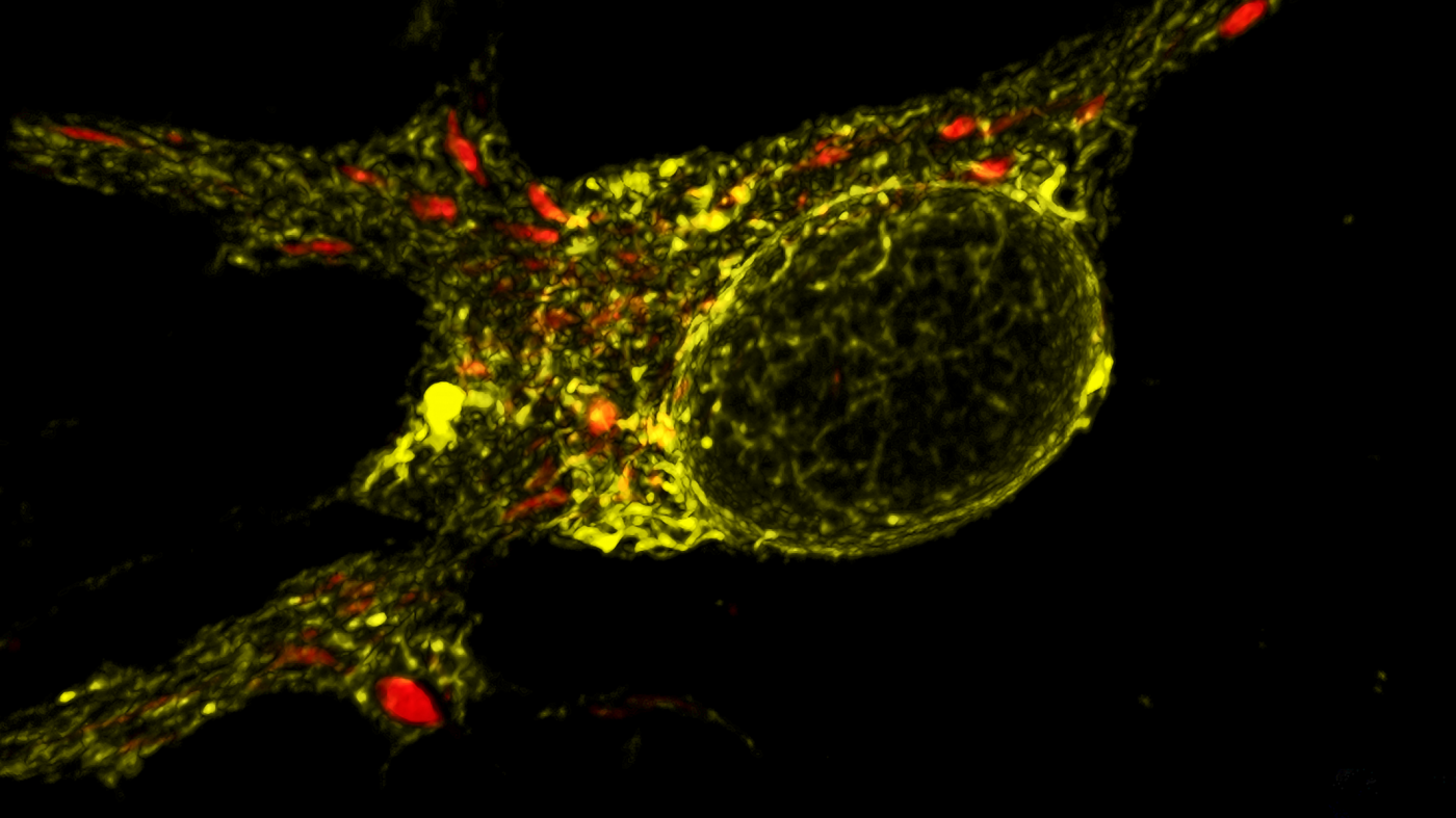
The new Zeiss LSM880 confocal microscope with Airyscan allows researchers to see tiny cellular components more clearly, like these mitochondria shown in red. [Image: Huihui Li, Gladstone Institutes]
In a small, dark room, four microscopes whir and hum while seven monitors glow with fluorescent images of biological specimens. A video of a mouse embryo plays on one of the screens, its beating heart and expanding and contracting lungs shine brightly in green and blue. Next to it, a spinal cord highlighted in orange and pink rotates on another monitor.
In just one year, the Histology and Light Microscopy Core at the Gladstone Institutes has gone from a sparsely equipped imaging center to one of the most technologically advanced facilities on the west coast. During that time, the core acquired three new microscopes that improve the attainable size, speed, and sensitivity of its imaging capabilities. Now researchers can image live cells, organs, and animal embryos in ways that were previously out of reach.
“When I came to Gladstone a little over a year ago, the core lacked many modern innovations in microscopy, particularly in resolution, speed, and size. It actually limited the research scientists could do,” said Meredith Calvert, PhD, director of the core. “With these upgrades, we can now take almost any sample and create any type of image from it, and at much faster speeds.”
Imaging Bigger, Not Smaller
The first addition to the core, the Zeiss Lightsheet Z.1, allows scientists to image much larger samples, such as mouse embryos, whole organs, or organoids—which mimic organ structure and function—created from stem cells.
In this microscope, two parallel sheets of light penetrate the sample, which is suspended in a column. A third lens takes 1000 pictures per minute, 10 times faster than standard confocal microscopes. Because the microscope also acts as an incubator, the samples can survive for several days, allowing scientists to track how live cells or tissues change in real time.
Todd McDevitt, PhD, who helped acquire the microscope, is using the Lightsheet to track the integration of neurons created from stem cells into damaged mouse spinal cords. The microscope can rotate the sample and take images from every angle, allowing McDevitt and his team to follow the growth of the cells and see where they repopulate the spinal cord. Another early adopter of the technology is Benoit Bruneau, PhD, whose lab members are using the Lightsheet to capture the origins of congenital heart defects precisely when and where they occur in a mouse embryo.
Improving Resolution
The second microscope added to the core was the Zeiss LSM 880 confocal microscope with Airyscan, which replaced Gladstone’s original confocal microscope. With significant advances in technology occurring every couple of years, the LSM 880 is a much-needed update. This highly sensitive microscope allows scientists to obtain much sharper three-dimensional images, even in very low light.
With the addition of Airyscan, the LSM 880 is also the first super-resolution microscope in the core. Previously, all of the microscopes at Gladstone had a resolution limit of 250 nanometers (the diffraction limit of light). Airyscan, a technology based on breakthroughs that won the Nobel Prize in Chemistry in 2014, expands that limit to 120 nanometers, the size of a single small bacterium. The improved resolution helps researchers to, among other things, clearly define much smaller objects. This capability is particularly important when studying whether two cellular entities interact or just sit next to one another.
The new technology has already advanced Gladstone science. For example, Ken Nakamura, MD, PhD, used the LSM 880 to make new discoveries about how mitochondria—the energy centers of the cell—relate to neurodegeneration.
Optimizing for Speed
The third piece of new equipment dramatically increases the core’s speed capabilities. The Zeiss Cell Observer Spinning Disc acquires images at 30 frames per second, capturing high-speed biological events, such as calcium signaling, in real time.
This technology fills needs in multiple areas. Li Gan, PhD, hopes to use the microscope to image firing cells in live brain slices, while Leor Weinberger, PhD, is interested in studying the processes by which a virus enters a cell in real time.
Celebrating the Upgrades
The Microscopy Core showcased its new capabilities with an open house on July 18, 2016. The upgrades to the core were made possible by the efforts of Gladstone investigators McDevitt, Gan, Lennart Mucke, MD, and Deepak Srivastava, MD, and a long-standing relationship with Zeiss, an international leader in microscopy technology.
The facility now houses a total of 12 microscopes and a support team that includes Calvert, two full-time histologists, and two summer interns. The core serves more than 70 percent of the laboratories at Gladstone in addition to 30 groups from outside the Institutes.
Looking to the future, Calvert said, “I’d like to move towards a full-service core, where scientists can drop off a sample with us and we return to them fully processed and analyzed image data. As the machinery becomes more and more advanced, this will be the best way to optimize the technology and enhance Gladstone’s research. Ultimately, we think this equipment will lead to more ground-breaking discoveries and help move the needle towards better treatments for disease."

